Shimane University Faculty of Medicine
ISSN :0386-5959(in print)
ISSN :2433-2410(online)

These article are licensed under a Creative Commons Attribution-NonCommercial-NoDerivatives 4.0 International License.
number of downloads : ?
Use this link to cite this item : https://ir.lib.shimane-u.ac.jp/34556
Shimane Journal of Medical Science 5 2
1981-12-01 発行
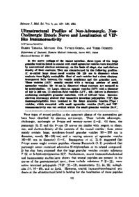
Ultrastructural Profiles of Non-Adrenergic, Non- Cholinergic Enteric Nerve and Localization of VIP-like Immunoreactivity
Tanaka, Osamu
Oki, Mitsuru
Gonda, Tatsuo
Domoto, Tokio
File
Description
In the nerve endings of the canine intestine, three types of the large granular vesicles found to coexist with small agranular vesicles were identified by conventional electron microscopy, on the basis of shape, size and electron density of their contents. They are characterized by the following profiles : (1) so-called large dense-cored vesicles (80-120 nm in diameter) whose contents were highly osmiophilic. Most of such vesicles had a clear-electron-transparent halo between the vesicle membrane and the granular core. These vesicles (LGV) usually coexist with a varying number of small agranular vesicles (50-70 nm in diameter) whose contents are assumed to be acetylcholine. (2) Large, electron-opaque vesicles (LOV) with a diameter of 140 to 200 nm. (3) electron-faint vesicles (LFV; 100-140nm in diameter) containing osmiophilic granular materials, with or without halos. Immunoelectron microscopy showed that vasoactive intestinal polypeptide (VIP)-like immunoprecipitates were localized to the large granular vesicles (Type 1 vesicles) which coexisted with small agranular vesicles (SAV), and VIP-immunoreactivity was not evident within the small granular vesicles (SGV).
About This Article
Other Article
PP. 123 - 129
PP. 109 - 122
PP. 130 - 135