Shimane University Faculty of Medicine
ISSN :0386-5959(in print)
ISSN :2433-2410(online)

These article are licensed under a Creative Commons Attribution-NonCommercial-NoDerivatives 4.0 International License.
number of downloads : ?
Use this link to cite this item : https://ir.lib.shimane-u.ac.jp/28603
Shimane Journal of Medical Science 30 1
2013-11-01 発行
Removal of Submucosal Foreign Body of the Hypopharynx Using
Noriaki, 70262071
Shimizu, Kanako
Yukie, 70330159
Mitsuhiro
Akemichi
File
Description
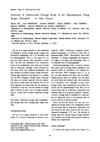
The use of an image intensifier is well established in orthopedics, trauma, urology, general surgery and intraarterial angiography, but is an unfamiliar tool for otolaryngologists. This is a case report of a 72 year-old female patient, who swallowed a metal wire. The wire was embedded in the submucosal tissue of the hypopharynx, and could not be found with an endoscope, although it was visualized by soft tissue X-ray and computed tomography, and was removed successfully using an image intensifier.
Foreign body impaction in the pharynx is a common case for ENT emergency. Foreign bodies, especially fish bones, are usually impacted into the oropharynx,
and are easily found and removed. The case of deep impaction of foreign body into the submucosa of pharynx is very rare, and the removal of foreign body is difficult in such a case. An image intensifier may be a useful tool to remove submucosal foreign body.
About This Article
Other Article
PP. 33 - 36