Faculty of Agriculture, Shimane University
ISSN:0370-940X
number of downloads : ?
Use this link to cite this item : https://ir.lib.shimane-u.ac.jp/3739
Bulletin of the Faculty of Agriculture, Shimane University 4
1970-12-15 発行
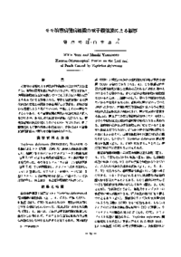
モモ縮葉病罹病組織の電子顕微鏡による観察
Electron-Microscopical Stusies on the LeaL-curl of Peach Caused by Taphrina deformans
Nozu, Mikio
Yamamoto, Masaki
File
Description
Ultra-thin sections of the leaf curl tissue of peach (Prunus persica SIEB. et ZUCC. var. vulgaris MAXIM.) infected with Taphrina deformans (BERKELEY) TULASNE Were investigated by means of an electron-microscope.
Pathogens (Figs. 1-4. H) were recognized in inter-cellular spaces (Figs. 1 -4. CS), middle lamella (Fig. 2) and cell walls (Fig. 3. CW) of the suscept tissue. Cell wall and cell membrane of the suscept cell was not invaginated and also haustorial structure was not observed. Suscept tissue consists of hypertrophied cells and young cells. Hypertrophied cell had a central vacuole (Figs. 1, 4, 5, 6, 7. V) and a thin peripheral layer of cytoplasm. Sometimes, nucleus had two nucleoli (Fig. 5). Starch grains (S) and rarely, lamellar structures were recognized in the chloroplasts (Figs. 6, 7. CH) of the hypertrophied cells. On the other hand, young cell had not a central vacuole. Golgi bodies (Figs. 8, 9. G), finger-print like pattern (Figs. 9, 15. FP) and various plastids, i. e. proplastid (P) and chloroplasts (CH) were found in the cytoplasm. Osmiophilic granules (OS) were observed inall plastids (P, CH) and sometimes, vacant spaces (VS) were found in the chloroplasts (Figs. 13, 14).
Pathogens (Figs. 1-4. H) were recognized in inter-cellular spaces (Figs. 1 -4. CS), middle lamella (Fig. 2) and cell walls (Fig. 3. CW) of the suscept tissue. Cell wall and cell membrane of the suscept cell was not invaginated and also haustorial structure was not observed. Suscept tissue consists of hypertrophied cells and young cells. Hypertrophied cell had a central vacuole (Figs. 1, 4, 5, 6, 7. V) and a thin peripheral layer of cytoplasm. Sometimes, nucleus had two nucleoli (Fig. 5). Starch grains (S) and rarely, lamellar structures were recognized in the chloroplasts (Figs. 6, 7. CH) of the hypertrophied cells. On the other hand, young cell had not a central vacuole. Golgi bodies (Figs. 8, 9. G), finger-print like pattern (Figs. 9, 15. FP) and various plastids, i. e. proplastid (P) and chloroplasts (CH) were found in the cytoplasm. Osmiophilic granules (OS) were observed inall plastids (P, CH) and sometimes, vacant spaces (VS) were found in the chloroplasts (Figs. 13, 14).
About This Article
Other Article
PP. 1 - 5
PP. 44 - 49
PP. 110 - 112
PP. 129 - 133
PP. 146 - 152
PP. 169 - 174
PP. 190 - 197
PP. 212 - 221