Faculty of Agriculture, Shimane University
ISSN:0370-940X
number of downloads : ?
Use this link to cite this item : https://ir.lib.shimane-u.ac.jp/1586
Bulletin of the Faculty of Agriculture, Shimane University 10
1976-12-15 発行
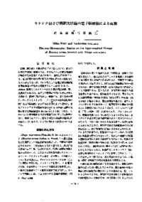
カラシナ白さび病肥大組織の電子顕微鏡による観察
Electron-Microscopical Studies on the Hypertrophied Tissues of Brassica cerma Infected with Albugo macrospora
Nozu, Mikio
Ishihara, Yoshimitsu
File
Description
Ultra-thin sections of the hypertrophied stem-tissues of Brassica cernua infected by Albugo macrospora were studied under an electron-microscope. Hypertrophied cell had a large vacuole and a thin peripheral layer of cytoplasmic area. Sometimes plasmolysis was observed but the disintegration of double membrane was not seen in nuclei, chloroplasts and mitochondria. Fungal hyphae were recognized in inter-cellular spaces and inside of the cell of suscept tissue.
Although the tips of hyphae were recognized in the suscept cell, the hyphae(hausto rium) were always surrounded by an invaginated cytoplasm. In the haustorial cells, large mitochondria, endoplasmic leticula and vacuole were found.
Although the tips of hyphae were recognized in the suscept cell, the hyphae(hausto rium) were always surrounded by an invaginated cytoplasm. In the haustorial cells, large mitochondria, endoplasmic leticula and vacuole were found.
About This Article
Other Article
PP. 39 - 42
PP. 66 - 73
PP. 125 - 129
PP. 135 - 140
PP. 160 - 172