| File | |
| Title |
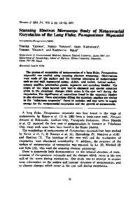
Scanning Electron Microscope Study of Metacercarial Excystation of the Lung Fluke, Paragonimus Miyazakii
|
| Creator |
Yamane Yosuke
Yoshida Nobuo
Nakagawa Akio
Makino Yumiko
Hirai Kazumitsu
|
| Source Title |
Shimane journal of medical science
|
| Volume | 3 |
| Issue | 1 |
| Start Page | 13 |
| End Page | 22 |
| Journal Identifire |
ISSN 03865959
EISSN 24332410
|
| Descriptions |
Abstract
The process of excystation of metacercariae of a lung fluke, Paragonimus miyazakii was studied using scanning electron microscopy. Observations were made of the surface and the internal structures of metacercariae such as cyst wall, tegumental spines, stylets, oral sucker, ventral sucker, sensory papillae, penetration glands, tegument and excretory bladder. The origin of the triple-layered cyst wall is discussed and special attention given to the structural changes which occur in the cyst wall during the excystation. The significance of concretions found in the excretory bladder is also discussed. These concretions filling the excretory papillae are similar to the calcareous corpuscles found in cestodes, and may serve to supply energy for the metacercarial excystation and the growth of metacercaria.
|
| Subjects |
excystation
Paragonimus
SEM
|
| Language |
eng
|
| Resource Type | departmental bulletin paper |
| Publisher |
Shimane Medical University
|
| Date of Issued | 1979-06-01 |
| Publish Type | Version of Record |
| Access Rights | open access |
| Relation |
[NCID]
AA00841586
|