Shimane University Faculty of Medicine
ISSN :0386-5959(in print)
ISSN :2433-2410(online)

These article are licensed under a Creative Commons Attribution-NonCommercial-NoDerivatives 4.0 International License.
number of downloads : ?
Use this link to cite this item : https://ir.lib.shimane-u.ac.jp/34667
Shimane Journal of Medical Science 16 1
1998-06-01 発行
Development of the hip joint in human embryos and fetuses
File
Description
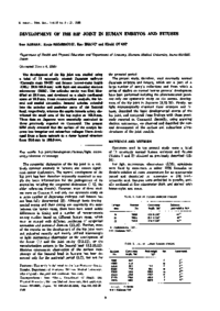
The development of the hip joint was studied using a total of 11 externally normal Japanese embryos (Carnegie stage 19-22) and fetuses (crown-rump length (CRL) 29.8-168.0mm) with light and scanning electron microscopy (SEM). The articular cavity was first identified at 29.8mm, and developed as a single continuous space at 78.0mm. From reconstruction analysis, the lateral and medial circumflex femoral arteries extended into the anterior and posterior parts of the femoral head, respectively, whereas the capitis femoris artery distributed the small area of the top region at 168.0mm. These data on Japanese were essentially equivalent to those previously reported on Caucasoid. The present SEM study revealed that the surface of the condyle became less irregular and subsurface collagen fibers developed from a loose network to a dense layered structure from 58.0mm to 168.0mm.
About This Article
NCID
AA00841586
Other Article
PP. 15 - 18